This article forms part of an educational supplement sponsored by Shockwave Medical. Explore the full Shockwave E8 and Javelin series here.

The Shockwave E8 catheter “significantly broadens the therapeutic landscape” for patients with diffuse below-the-knee (BTK) and below-the-ankle (BTA) disease. This is according to Marta Lobato and August Ysa (Barakaldo, Spain), who here present a case in which the Shockwave E8 played a key role in treatment success.
A 76-year-old male presented to our department with signs of chronic limb-threatening ischaemia (CLTI) and a rapidly worsening diabetic foot infection following prior debridement at another centre. He was a former smoker with a history of hypertension, diabetes mellitus, dyslipidaemia and chronic kidney disease. He was under regular treatment with metformin, enalapril, acetylsalicylic acid (ASA) and atorvastatin.
Initial presentation and clinical evaluation
The patient was referred urgently due to the appearance of an infected forefoot ulcer with signs of local sepsis. He was febrile, with a white blood cell count of 15,000cells/mm³, and had no palpable pedal pulses. The patient also had acute renal failure, further complicating his clinical status. The ulcer was extensive, with surrounding cellulitis and necrotic tissue. A bedside ankle– brachial index (ABI) was non-compressible, likely due to arterial calcification, and transcutaneous oxygen pressure (TcPO2) on the dorsum of the foot was critically low at 12mmHg.
The lesion was classified using the Wound, Ischemia, and foot Infection (WIfI) system as stage 3-3-3, reflecting high risk of limb loss. An urgent surgical debridement was performed, followed by initiation of broadspectrum intravenous antibiotics.
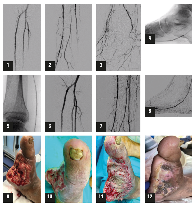
Imaging and decision-making Subsequent diagnostic angiography revealed severe multilevel calcific arterial disease, with diffuse narrowing and dense calcification throughout the tibial vessels. The anterior tibial artery (ATA) was patent in its proximal and mid segments with significant lesions, but showed distal occlusion with no reconstitution at the level of the foot. The posterior tibial artery (PTA) exhibited multiple stenoses with heavy calcification, distal third occlusion, and reconstitution at the level of the plantar artery (Figures 1–3). Based on these angiographic findings, a decision was made to proceed with an endovascular-first strategy, with the main goal of re-establishing direct in-line flow to the ischaemic wound site via the PTA.
Endovascular procedure: Leveraging Shockwave E8
An antegrade femoral approach under ultrasound guidance was perfromed. A long sheath was advanced and positioned in the popliteal artery to provide stable support. After crossing the PTA occlusion with a 0.014” guidewire, the decision was made to use the Shockwave E8 catheter as the primary tool for vessel preparation. This decision was based on several key factors. The lesion’s morphology showed extensive concentric calcification, which limited the effectiveness of standard angioplasty and carried a high risk of dissection or recoil. The Shockwave E8 catheter’s enhanced pushability and trackability made it ideal for navigating the tortuous, narrow tibial anatomy. Furthermore, the longer integrated balloon of the Shockwave E8 allowed the entire diseased segment to be treated with a single device, reducing the need for multiple exchanges and improving procedural efficiency.
The Shockwave E8 catheter was advanced across the lesion and activated, delivering 400 pulses at low inflation pressures of 2–4atm. This achieved improved compliance through modification of the calcified plaque and enabled subsequent balloon expansion without complications. The procedure was completed with a non-compliant balloon angioplasty to optimise luminal gain (Figures 4–5).
Results and postoperative course
Post-Shockwave E8 angiography showed excellent luminal gain, with no flowlimiting dissections or residual stenosis requiring further intervention. The final result demonstrated significantly increased flow through the PTA into the plantar arch, confirming technical success (Figures 6–8).
Within hours of the procedure, the posterior tibial pulse returned, and follow-up TcPO2 increased dramatically to 61mmHg, correlating with successful reperfusion. The patient continued antibiotic therapy (ciprofloxacin and clindamycin) and underwent vacuum-assisted closure (VAC) therapy to promote wound healing.
He was discharged 10 days later, under close home-care monitoring. A split-thickness skin graft was performed in the following weeks, and progressive wound closure was observed. By the end of follow-up, the wound had completely healed, and the patient had recovered full ambulatory function (Figures 9–12).
Discussion
This case highlights the increasing role of Shockwave intravascular lithotripsy (IVL) as a frontline tool in the treatment of complex, calcified BTK disease, particularly in patients with CLTI and limited revascularisation options. Calcium modification is essential in these scenarios, not only to achieve immediate luminal gain but also to minimise trauma and avoid stenting, particularly in the distal BTK and BTA segments, where vessels are small, mobile, and at high risk of fracture or occlusion.
While the Shockwave S4 catheter has played a central role for IVL in small BTK vessels, in our practice the introduction of the Shockwave E8 offers several practical and clinical advantages. The device has enhanced pushability and flexibility, allowing it to cross tight, calcified, and tortuous segments more efficiently, particularly in distal tibial and foot territories. Its extended treatment segment permits operators to treat long, calcified lesions with a single device, improving efficiency, reducing procedure time, and minimising the number of catheter exchanges. Treating a full-length tibial lesion with one catheter also reduces the overall procedural cost and simplifies logistics. In addition, the uniform energy delivery and low-pressure inflation reduce barotrauma, improving safety and reducing postprocedural complications.
In conclusion, the Shockwave E8 catheter significantly broadens the therapeutic landscape for patients with diffuse BTK and BTA disease by enabling safe, effective, and comprehensive treatment of long, calcified arterial segments. In the setting of CLTI, preserving native arterial anatomy and maintaining future options is crucial. The Shockwave E8 represents a true paradigm shift in the management of infrapopliteal calcification and represents a new valuable tool in BTK and BTA endovascular care.
Case images
Marta Lobato and August Ysa are both vascular and endovascular surgeons in the BTK-BTA Unit at Hospital de Cruces in Barakaldo, Spain and paid Shockwave Medical consultants. The views expressed in this article are those of the physicians and may not reflect the views of Shockwave Medical.


