A multidisciplinary team of researchers at University of California (UC) San Diego (San Diego, USA) report that photoplethysmography (PPG) coupled with artificial intelligence (AI) can detect peripheral arterial disease (PAD) with a high degree of accuracy. The technique could be used as a digital biomarker to quickly and non-invasively screen for the condition, potentially leading to earlier diagnosis and treatment. The study was recently published in npj Digital Medicine.
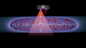
“PPG works by shining a light into tissue, in our case, the toe,” said co-first author Ava J Fascetti, a PhD student in the lab of senior author Edward J Wang. “A photosensor measures how much light is reflected back, allowing us to detect tiny changes in blood volume: what we call the PPG signal.”
A UC San Diego press release notes that the idea for the study came about when co-first author Mattheus Ramsis had a conversation with co-author Elsie G Ross and learned that PPG recordings are routinely taken on the toe at the same time patients undergo ABI testing.
“The lightbulb went off for me at that moment,” said Ramsis.
He enlisted fourth-year medical student Mustafa H Naguib to assemble a dataset of over 10,000 toe PPG recordings collected from more than 3,500 patients who underwent ankle‑brachial index (ABI) tests at UC San Diego Health between 2020 and 2025. The team then extracted 78 PPG features that were significantly correlated with the patients’ ABI results. From there, they developed a machine learning model to detect whether a patient had PAD based solely on those PPG features.
“When we built the model using only the PPG data, it demonstrated strong performance in distinguishing patients with PAD (defined by an abnormal ABI) from those without the disease, correctly distinguishing PAD cases approximately 83% of the time, compared with the roughly 60–65% performance typically achieved using traditional clinical risk-factor assessment alone,” said Ramsis. Adding a patient’s smoking status improved the model by another 2%. “Importantly, the signal reflects physiologic blood flow changes in the toes, providing information beyond conventional clinical evaluation.”
What’s more, the model performed similarly across Black, Hispanic and white patients, as well as on data from two separate UC San Diego Health medical campuses, including patients with coronary artery disease, diabetes and end-stage renal disease.
“These findings represent an initial step toward identifying an accessible, physiologically grounded digital biomarker associated with PAD, and lay the foundation for prospective studies to evaluate performance across clinical workflows and reference standards,” the authors write in npj Digital Medicine.
Ramsis says the next step is to validate the approach across multiple PPG capturing devices including smartphones, pulse‑oximeters and wearable technologies to determine whether the model will hold up in real‑world settings. While there are no plans to replace ABI testing with PPG screening for now, he thinks it could complement the more established method.
“An ABI takes 15-30 minutes once the patient is in the clinic, whereas a PPG screen takes only a few seconds,” he said. “Because 95% of people own a smartphone or some advanced device, we can potentially bypass the transportation, financial and institutional barriers that currently limit access to ABI testing.”
A quick PPG screen could be used as a point‑of‑care triage in the clinic, or high-risk patients could even screen themselves on their phone with a smartphone app, prompting earlier referral before a major adverse limb event occurs.
“If we can catch PAD early enough to prevent a limb amputation, that would be the ultimate impact: preserving limb function, reducing mortality and addressing barriers in underserved populations,” said Ramsis.
“We envision this work as an early example of how scalable biosignal infrastructure can support the development of next-generation digital diagnostics across the health system,” added Ramsis. “The physiologic data collected through studies like this will help create the foundation for future biosignal-based research, translation and clinical implementation.”
The study was funded, in part, by the American College of Cardiology Foundation, Accreditation Foundation Committee.