
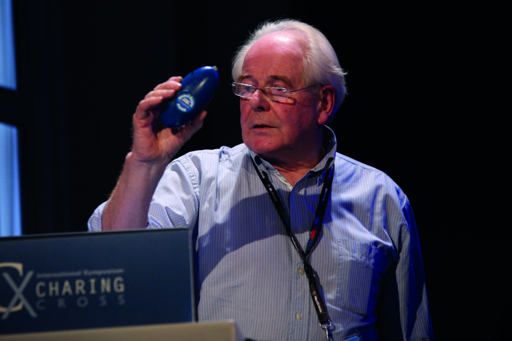
BlueDop (BlueDop Medical) is a wireless egg-shaped device that converts a Doppler signal to a readable pressure measurement. Vascular News spoke to its inventor and BlueDop Medical managing director, David King, who explained why the device—winner of the 2014 CX Innovation Showcase Dragons’ Den—offers an alternative to traditional ankle brachial index assessment.
Can you briefly explain what BlueDop is?
BlueDop is a smart Doppler device which targets the shortcomings of traditional ankle brachial index in the assessment of lower limb arterial disease. It comes in two parts; a hand-held Doppler ultrasound device we call “the egg” and a tablet computer display we call “the brain”. The egg is completely sealed in a silicone rubber casing and transmits directional Doppler audio via Bluetooth technology to the brain, recharged using inductive technology similar to your electric toothbrush. The brain instantaneously analyses and displays the directional Doppler spectrum to allow the operator to adjust for best blood flow signals from the tibial arteries. Now here is the clever bit; the brain does four things that are unique to BlueDop technology. It rejects non-physiological flow signals, estimates mean perfusion pressure in the foot without using a leg cuff, detects the presence of significant arterial disease and indicates the presence of critical limb ischaemia using a pressure based measure of vascular reserve.
Why is the current technology not adequate for peripheral artery disease triage?
Conventional ankle brachial index does not work well in the diabetic foot, in swollen and ulcerated limbs and in the chair-bound patient. These are the very patients who urgently require triaging either at home, in the physician’s office or in a tissue viability clinic. Provided an adequate blood flow signal is detected, we believe BlueDop’s performance will not be affected by arterial calcification, limb swelling, patient positioning, the presence of ulceration or extensive wound dressings.
How does BlueDop calculate mean arterial pressure?
We know from our research that in the maximally exercised state the shape of the instantaneous flow wave supplying an organ such as the calf muscle is identical to the shape of the driving pressure wave created in the ascending aorta. This occurs because the vascular system defaults to the matched state when stressed. So why is this important? Theoretically this is where maximum hydraulic power is dissipated in the organ under study. Moreover at this point the mean organ perfusion pressure is exactly half that of the ascending aorta—no “ifs or buts”, it just comes out of the science. A little lateral thinking and a further big inventive step permits estimation of perfusion pressure over what we believe to be the complete physiological and pathological range, provided we supply a reference pressure representative of that driving pressure waveform. This can be obtained with a simple automatic blood pressure machine on the arm.
What have preliminary studies involving BlueDop shown?
Direct intra-arterial pressure correlations have been carried out in an in vivo canine model over the full physiological pressure range down to one half aortic mean pressure. The pressure from flow correlated well with an art line pressure measurement. In additional preliminary studies we have compared the disease detection mode of our system to duplex. Early results have been promising. All experiments described here conformed to national ethical standards.
Perfusion pressure, ankle brachial index and toe brachial index are blood flow dependant variables, particularly in the presence of arterial disease, so no specific pressure or pressure ratio suggests itself as an indicator of significant arterial disease. BlueDop uses a bistable “disease/no disease” indicator we call the C notch to detect the presence of significant peripheral arterial disease in the resting patient. The magnitude and timing of the C notch differentiates it from the dichrotic notch (or D notch). The C notch is generated only in the absence of significant arterial disease and so the absence of the C notch indicates the presence of significant arterial disease. We have automated the C notch detection with a proprietary process because the computer is better than we are at picking up the subtle timing and magnitude of information held in the Doppler spectrum.
What is the next step for BlueDop?
The next step for BlueDop is to obtain the requisite regulatory approvals that are required before we can make the device and its technology available to clinicians, nurses and podiatrists. We are hoping to obtain a diagnostic code in the USA and that will require us to carry out an investigational device exemption. We are lucky in having some really experienced marketing and regulatory consultants as part of our US team. CE approval is the next step for the European market and we are in the process right now. We have a number of clinical groups in Europe and the USA who wish to carry out independent, double-blinded clinical trials and both sides are keen to get going on this. Both the European and US clinical trials will contribute to the US process.
Is there anything else we have not covered that you would like to mention?
Just to point out that our pressure from flow algorithm is applicable in arteries almost anywhere in the body so that a whole bunch of additional applications become possible. We are looking at dialysis fistula monitoring and functional stenosis testing using exercise and these are very promising, but we welcome any “cool” new ideas if they come along.


