Tilo Kölbel (Hamburg, Germany; moderator) is joined by Geert Willem Schurink (Maastricht, Netherlands) and Joost van Herwaarden (Utrecht, Netherlands) in this Vascular News roundtable, titled Bringing 3D device guidance into the hybrid lab with Fiber Optic RealShape (FORS) technology.
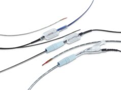
FORS technology (Philips) was first introduced into clinical practice in 2018. Since then more than 130 cases–the majority aortic but some peripheral–have been performed with the technology across three European centres. The trio of experts share case examples that highlight some of the “most interesting features” of FORS and discuss how the technology can help improve clinical practice.
Van Herwaarden presents some peripheral cases using FORS and lists some of the key features including its ability to visualise a wire and catheter in 3D and in real time, using light rather than X-Ray, and the fact that it can be used in bi-plane mode.
Kölbel talks about the learning curve with the technology and notes that although the radiation reduction is “significant”, the workflow improvements are perhaps “even more important”. He goes on to state that “the lateral views have the biggest impact on radiation”, adding, “I use the angulations less and that saves time and radiation”.
Schurink presents a case using the bi-plane mode during cannulation of a left renal artery with a steerable sheath, noting that the technology allows you to “really aim at your target vessel”. Schurink further outlines how development of FORS could lead to it being used in a growing number of interventional procedures, explaining that “we are now at the very beginning of this journey but I think there is a lot to gain in the endovascular world”.
For more information on FORS technology, click here.
This video is sponsored by Philips.


