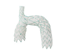
 At the 2017 Charing Cross Symposium (25–28 April, London, UK), speakers and panellists discussed the challenges of endovascular aortic arch procedures and their potential impact on acute stroke. Looking at both the causes and potential solutions for this problem, those involved noted that while understanding has increased, significant obstructions and risks still remain.
Stéphan Haulon (Lille, France) spoke on the concern over cerebral embolisation risk caused by endovascular procedures in the ascending aorta and the aortic arch, specifically with the use of branched endografts, and potential steps to reduce such a risk. With this approach, Haulon explained, “Obviously crossing the arch and aortic valve with the wire poses a risk right from the beginning. Patient selection is important as having a patient with a shaggy aorta in the arch is an absolute contraindication. To avoid stroke at that time of the procedure, centre line navigation (navigating in the middle of the aortic lumen) is mandatory. That can be done by using fusion, steerable sheaths or robotic guidance.” Pushing a wire into the left ventricle can also offer a risk of stroke, Haulon said, creating left ventricular false aneurysms. Positioning a sheath in the supra-aortic trunk, especially in the right common carotid artery, increases stroke risk, which is why Haulon and colleagues maintain aggressive anticoagulation in these cases. A third concern is emboli caused by contact of the endograft delivery system with the greater curvature of the arch, the risk of which can be lessened by reducing the profile of the introducer, navigating over a stiff, double-curved wire placed into the left ventricle, and using a transapical or trans-septal approach.
Utilising an endograft designed to position catheters further from the aortic wall is also important to consider, Haulon said, as is adequate endograft flushing—with both saline and CO2—to avoid any risk of air embolism during deployment. Finally, supra-aortic clamping and the use of protective filters could also be considered as stroke risk reducers, although Haulon noted that “there is still a lot of research to be performed” on their use.
Tilo Kölbel (Hamburg, Germany) spent part of his presentation looking at one element of Haulon’s presentation—the dangers of air embolism—sharing his team’s research with the audience. “The mechanism of stroke is still unclear,” Kölbel explained, “and we have, so far, mainly considered solid emboli as the main cause of stroke. Air embolism is another potential cause of stroke.” Even when the instructions for use are followed, air can still remain in the endograft before deployment, Kölbel said. This air can then enter the brain causing stroke, and, in some cases, accounting for up to 90% of high-intensity transient signals recorded when using intraoperative trans-cranial Doppler, the majority of which occur during device deployment. “Saline flushing is not enough to fully clear stent grafts of air before deployment,” said Kölbel, showing videos taken in Hamburg of air escaping from stent grafts as they are deployed post-flushing, and even though the amount of air may be small, “if released into sensitive areas it can do damage.”
“We may have been barking up the wrong tree,” Kölbel suggested. “I am sure that the cause of stroke is multi-factorial, and air embolism may play a significant role in this,” he concluded.
Heinz Jacob (Duisburg, Germany) then addressed delegates on avoiding embolisation in open procedures. “Stroke is a major concern in open arch surgery,” Jacob said. The armamentarium available to combat this concern includes epiaortic scanning, modified cannulation techniques, perfusion management, CO2 insufflation, and near-infrared spectroscopy.
Epiaortic scanning is helpful “because it enables visualisation of intramural/intraluminal lesions, ulcerous lesions and floating structures,” Jacob said. This may influence decisions about the site of arterial cannulation, the level of aortic cross-clamping, secondary clamping, or even whether to clamp at all.
With perfusion management, the goal is continuous, uninterrupted flow to the brain with, whenever possible, all three head vessels. At Jacob’s centre, the team use whole body selective perfusion, using a venous line, an arterial line, and three suction devices. The method uses a separate circuit for selective perfusion of the left subclavian/ axillary artery and the downstream aorta. The patient is cooled to 28 degrees at the bladder, the blood cooled to 22 degrees for the arch and into brain, and then increased to 28 degrees once the anastomosis has been done for the lower body.
Central cannulation is used to access the true lumen, although this can often be difficult, and easy access “is rare”. In difficult cases, Jacob and colleagues use open vision aortic cannulation, in which the aorta is opened up once blood pressure is lowered, the sac is cut into and the cannula introduced. In cases of true lumen collapse, en bloc reimplantation is used.
Rodney White (Long Beach, USA) shared his centre’s experience, telling delegates that increased stroke incidence occurs with any procedure instrumenting the ascending aorta and arch, and that new technologies—including transcranial Doppler, intravascular ultrasound and transoesophageal echocardiography—are now providing incremental identification and improving care of stroke. He also suggested that new anticoagulation schemes are needed to provide peri-procedural protection while preventing bleeding.
Michael Dake’s (Stanford, USA) presentation focused on the risks associated with the use of branched endografts, explaining, “Our awareness of this issue and its relevance is increasingly critical as we take these technologies more towards proximal aortic use.” Dake looked at outcomes of a pivotal trial analysing the TAG thoracic branch endoprosthesis (Gore) for stroke risk, a trial for which Dake is a national co-principal investigator. He noted that preliminary results suggest that stroke risk varies depending on the zones being treated. For example, in zone 2, risk is relatively low (3.3%), while in zones 0/1 the risk is much higher (22.2%). This compares with a risk estimate of 7.4% for non-branched TEVAR in proximal descending thoracoabdominal aorta. Dake also told the audience that elderly patients, those with prior stroke, and those with high-grade arch or mobile atheroma were at increased risk. Acute outcome after post-TEVAR stroke is “dismal”, with a mortality rate of 33–57% (with rupture), while one-year survival after post-TEVAR stroke is “bleak”, at half the rate of survival of those patients with no stroke.
Alexandra Lansky (London, UK) touched on the potential value of cerebral protection devices, noting that their use has already been widely adopted for transcatheter aortic valve implantation (TAVI) procedures, and that they may also have a place in aortic arch procedures. Based on experience with TAVI, Lansky told the audience that the timing of a stroke is very important, with the most severe events occurring within one day of the start of the procedure. Lansky also suggested that “stroke rates are under-reported,” and that all endovascular cardiovascular procedures carry iatrogenic stroke risk and cause iatrogenic embolisation. Recent TAVI studies show that “every single patient has evidence of cerebral injury, caused by showers of large volume emboli.” In these patients there is a link between lesion and cognitive dysfunction, for example with a two-fold increase in dementia and a three-fold incidence of stroke; “It is a marker of subsequent stroke,” Lansky added.
Whether these embolisations are preventable is therefore a vital issue. Lansky referred to two US studies investigating the potential of filters to catch emboli. The SENTINEL US IDE trial—investigating the Sentinel system (Claret Medical)—met its primary safety endpoint, although it did not meet its superiority efficacy endpoint. “The key point with this trial is that, by histopathology, every single patient had emboli and debris captured in the filters,” Lansky said. The second trial analysed the TriGuard protection system (Keystone Heart). Lansky reported that the device showed a reduction in stroke rates and cerebral infarction.
Closing, Lansky stressed that “incidence of neurological injury really depends on how you define your endpoint, whether it is very severe or covert.”
Building on this, Gagandeep Grover (London, UK) presented her experience with cerebral embolic protection, telling the CX audience that the relation of so-called silent stroke to increased incidence of dementia, depression, neurocognitive decline and stroke, means that in the long-term, “Silent stroke is not so silent.” The Sentinel protection system—suitable for protection in zones 2–4—has had “encouraging” experience in TAVI. Grover and colleagues have deployed the device in 10 patients at their centre, with a mean age of 68 years and of which 70% were urgent cases. Early data show a 90% success rate, no device-related complications and no incidents of stroke. This was achieved with seven minutes added to the procedure time and a “negligible” radiation dose increase. Of the 19 filters, 18 caught embolic debris, with a median number of particles per filter of 937 and a per patient debris surface area of 0.54mm2. The most common types of debris were acute thrombus (95%), arterial wall (63%) and foreign material (32%). Distal filters captured more debris than the proximal filters.
Following the presentations, the speakers joined panel discussions to delve deeper into the topics raised in the main session and to give audience members the chance to ask questions about what they had just heard.
The importance of terminology
Gibbs asked whether placing patients in the Trendelenburg position might reduce embolism and the risk of stroke. Kölbel replied, “We have not tried that yet, and I do not think it is easy to change the geometry of the aortic arch significantly by tilting. I see that there may be an opportunity to tilt the table in the other direction; maybe we can direct the gas more towards the periphery. On the other hand, I think we should focus on avoiding putting air into patients, rather than directing it to less harmful areas.”
The panel also discussed the potential of rapid overdrive pacing when used to lower blood pressure, which Gibbs said “is great for positioning of the stent”. However, he questioned whether this method renders the brain vulnerable, because you are making it relatively ischaemic at the time that you are then subjecting it to the potential of embolisation. As Kölbel responded, “We know from other areas that we make the tissue vulnerable by ischaemia, and maybe we should use other ways of cardiac output reduction. In our experience, we do not need the complete stop of flow. Using inferior vena cava balloon occlusion with ascending stent grafts works quite well, reducing the flow, and possibly not damaging the brain as much.”
Haulon spoke of his centre’s experience with rapid overdrive pacing, noting that “what we have learned is that we have to keep it to the minimum also to reduce myocardial infarction. Some patients from the early experience did not recover from rapid pacing, so we now have techniques to reduce rapid pacing to 30 seconds, and also, we probably would need to start unsheathing the endograft inside the descending thoracic aorta to get air, if there is any left, to the lower limbs rather than the brain.”
Fiona Rohlffs (Hamburg, Germany) noted the importance of a team approach to cardiac output reduction, explaining, “It is not only us performing the procedure, we also need to discuss with the anaesthesiologist whether we need 100% oxygen during the procedure. It is really important that we have a team approach alongside the neurologists and anaesthesiologists.”
Further audience discussion analysed the importance of terminology and whether and how to differentiate between asymptomatic and symptomatic strokes. Anthony Rudd (London, UK) explained the World Health Organization definition, as “a clinical diagnosis of focal neurological deficit lasting more than 24 hours of presumed vascular origin.” However, he stressed that using this definition “does not mean that we should be ignoring the asymptomatic infarcts or haemorrhages. By definition, to pick up an asymptomatic thing you need to do an investigation with imaging, and as imaging gets more and more advanced, we are able to see smaller and smaller areas. We must not call these strokes, but on the other hand they can go on and progress to dementia and other things.”
Lansky supported this analysis, noting that “there is a lot of evidence linking these acutely asymptomatic strokes with subsequent neurological, cognitive dysfunction, so while we do not have that evidence in these procedures, I think that evidence does reside in other territories. It is important for us to keep that in mind and potentially to follow the patients and understand that longer term, and we do not do that.”
A question from the audience noted that the American Heart Association includes silent infarctions, to which Rudd responded, “If we are going to be using words in a different way, let us at least make sure that we define them very clearly.”
Discussing what he had gleaned from the session, Haulon said, “I think we have learned a lot this morning from all the speakers and there is great research going on. I am not going to talk about silent stroke, but it seems that it has an impact and pre- and postoperative magnetic resonance imaging (MRI) on this need to be analysed, not only in TEVAR but also for open surgery; we need to be careful not to compare apples and oranges in the literature.”
Haulon also suggested that an important future step could be a global study comparing all available filters. “We need to compare and we need to learn from everyone’s practice, and we need to do this independently. I think probably MRI is going to be the best way to compare the techniques,” he said.
Kölbel believes, “There are so many unknowns and we need to sort these things out; solid vs. gas, whether emboli protection devices actually help or are a risk of stroke, we need to learn whether we can transfer the knowledge from TAVI to TEVAR or whether this is a completely different area. What I think this session showed, mainly, is that there is a lot of work to do.”


