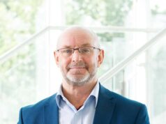
Wound care specialists can improve microcirculation by maximising “the overall vascular health of patients” It is the refrain William Ennis (University of Illinois, Chicago, USA) hears continually: “I cannot do anything about the microcirculation!” His swift response however, at the annual meeting of the American College of Wound Healing and Tissue Repair (ACWHTR 2019; 11–12 October, Chicago, USA) was that “you can do a lot by treating the patient.”
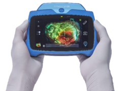
Ennis was speaking during a presentation entitled “Microcirculation: What, How, Why?”, in which he explained how imaging techniques can change what wound care specialists do, and offered an in-depth analysis of the slew of available devices.
He laid out some of the possibilities, pondering “whichever device you decide to use, when you come up with a positive finding, then what?”. Ennis, a professor of Surgery at the University of Illinois at Chicago, USA, and chief medical officer at wound care centre provider Healogics (Jacksonville, Florida, USA), added: “Can you change the microcirculation? Well, you can assess it before and after a procedure, before and after the of use energy-based modalities, or you could use an angiosome-based revascularisation procedure and then measure success or failure.
“I would argue the other thing we can do is maximise the overall vascular health of the patient. We can really work on getting patients to stop smoking and make sure to screen patients for sleep apnea, as well as making sure someone has been studied with an echo before we use compression bandaging and move a large amount of extravascular fluid centrally, to avoid tipping someone into congestive heart failure. We can work on the management of their hypertension, as that has an impact on the microcirculation. Also, we can analyse and try to lower central venous pressure for the same reasons.”
Ennis issued a caution on device selection. “What we are not going to do is tell you which of these devices to get,” he told delegates., before highlighting that there were a number of questions circulating in the wound village regarding this question. “I would rather ask you what question it is that you are trying to solve. For example, are you looking for a device that is a tool for the rapid screening of the overall arterial system? Then maybe you could use one of the devices to measure the microcirculation on the plantar surface of the foot, where pigmentation is not an impact, and that could act as a surrogate for the overall macrovascular status. This information could add to a patient’s history and physical exam and, perhaps, help you triage the patient for a vascular consultation.”
Other devices can add further information to the overall picture of wound healing, such as the SurroSense Rx sensory system insoles, which monitor both pressure and temperature.
“So think about the questions you are trying solve,” Ennis said. “That will help you to better understand what you want and which devices you need. It may end up, unfortunately, that you need more than one device. The type of device you need will also be dictated by the type of wounds you see in your clinic. for example, some clinics focus on diabetic foot ulcers (DFUs) only, while other see wounds of all aetiologies. Ultimately, this should lead to adequate macrovascular status with the end result being adequate delivery of oxygen and the formation of ATP. In the past, our group has used biopsy methods such as NMR spectroscopy to assess the ATP levels of wounds before and after procedures, but these tests are invasive, tedious and expensive”.
There is a “tremendous disconnect” between the testing and understanding of macro and microcirculation in the wound care space, Ennis said. “What I want to encourage people to do is to take a step back when you evaluate these types of imaging systems and think about the macro level first. When we start to think about the microcirculation however, we need to think about the vascular system as a whole going all the way to the level of the heart, as well as pulmonary hypertension, right ventricular failure, vena cava obstruction, lymphatic disease, and of course the arterial side of circulation”.
Ennis continued on the theme of complex systems theory when he stated that if you drink a cup of coffee in the morning prior to your microvascular testing, you would have increased peripheral vascular resistance and this could alter your results. One cigarette can result in 60 minutes of vasospasm and also produce erroneous testing when assessing the microcirculation.
Ennis then drew delegates back to the live surgical presentation from earlier in the conference, performed by his colleague Martin Borhani (University of Illinois, Chicago, USA). In this case, an endarterectomy procedure had been performed, resulting in increased blood flow to the calcaneus; on the live surgical case, a very robust granulation tissue was seen throughout the wound bed of the patient’s heel. A microvascular study was performed and demonstrated what appeared to be a very good blood flow to the granulation tissue.
Discussing the procedure, Ennis said: “Sometimes a patient like this will receive a skin graft and within 72 hours good pigmentation and engraftment is present, only for necrosis to begin five to six days after the procedure. By day 10, the graft might have been completely sloughed off.”
Ennis continued: “One of the possible reasons for this delayed skin graft failure could be the patient’s resumption of cigarette smoking in the immediate postoperative period, as these new, fragile vessels are susceptible to vasoconstriction from nicotine. Let us say, however, that you have a totally compliant patient who did not do any of the things that could harm the graft, and who states that they have kept their legs elevated. They then say: ‘I thought you guys told me I was ready for a skin graft; why did it not take?’
“By utilising devices that measure both the quantity and the quality of flow to the microcirculation, we might be able to gain an insight into when patients are ready for skin grafting and when the highest likelihood is that engraftment will proceed in an orderly fashion. It turns out that when providers have tremendous experience in this area, they are usually adept at determining skin graft take versus failure rates just by looking at the wound bed. That knowledge, however, is not present in many providers who only see a few wounds in any given year. The hope is that these devices will democratise knowledge and experience.”
Now, the field of wound care requires a piece of technology that steps into this void. “What we need, especially from our industry colleagues, is some type of device that helps to spread this knowledge and allows for a provider in Saint Elsewhere Hospital, who is new to wound care and has only been there for a year, to at least have some adjunctive tool to bring up their level of decision-making to someone who has seen a high volume of cases,” he said. “It does not mean that you do not need the experts in certain cases, but the technology can try to level the playing field by providing adjunctive assistance in wound healing decision-making.”


