This article is an advertorial by Lombard Medical.
The outcomes at five years of the Aorfix (Lombard Medical) PYTHAGORAS trial, published in the Journal of Vascular Surgery, provided the first long-term data from an endovascular aneurysm repair (EVAR) cohort to include a majority of highly angulated infrarenal necks. Vascular News spoke to investigator Mahmoud Malas about the trial and the implications of its findings.
What risks, difficulties or complications have typically been associated with EVAR in highly angled aortic necks?
Highly angled aortic necks (≥60° angulation between infrarenal aortic neck and the longitudinal axis of the aneurysm) increases EVAR complexity by making it difficult to introduce the delivery system and deploy the stent graft. It is also associated with several postoperative complications. More recent data have shown an association between difficult aortic anatomy and decreased long-term survival.
How does the Aorfix endograft design address these issues?
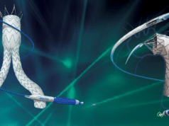
The Aorfix endograft is designed to be flexible and conformable to the vessel which makes it particularly suitable when proximal and/or distal landing zones are considerably angulated. It employs polyester graft material surrounded by independent nitinol rings, rather than Z stents, allowing the device to conform to the angulation of the native aorta without collapsing, and be resistant to kinking. Four pairs of hooks proximal on the graft are attached within an 8mm primary sealing ring to allow transrenal fixation. When implanted, the stent graft rings reform to a saddle or “fish mouth” shape, hugging the renal artery orifice and allowing the endograft to be placed pararenally. Moreover, the troughs align with the renal arteries juxtarenally and the peak extends suprarenally, providing optimum seal positioning and reducing risk of endoleaks.
How was the PYTHAGORAS trial designed?
The PYTHAGORAS trial is the first clinical trial designed to test the use of EVAR for highly angulated (≥60°) aortic necks. It is a multicentre (45 institutions), non-randomised regulatory study approved by the US FDA. The study enrolled 218 patients for an Aorfix procedure divided into two groups: standard aortic neck angulation (<60°) and highly angulated aortic necks. Patients were followed up for a total of five years. Postoperative imaging studies (CT scans and abdominal radiographs) were reviewed to assess all components of the primary efficacy endpoints including all-cause or aneurysm related mortality, endoleaks, rupture, reintervention and graft migration.
What were the results of the trial at five years, and what data did you find noteworthy?
I believe that the most interesting findings from the PYTHAGORAS study is that it is the first clinical trial to include patients with highly angulated aortic necks (69%), who would be deemed at high risk for EVAR with other endovascular aortic devices. The results are favourable and support the use of this on-label endovascular option. Freedom from all-cause mortality (69%) aneurysm-related mortality (96%), secondary intervention, and aneurysm rupture rates over five years were comparable with the Lifeline Registry of United States EVAR trials in normal anatomy and were found to be equivalent to other endografts used to treat normal anatomy. Moreover, the five-year sac expansion rates (12%) were lower than those of prior studies. The trial also reported no differences in the rate of type I/III or type II endoleaks or need for reinterventions with Aorfix among patients with standard-angle versus highly angulated aortic necks.
How do you think these results may impact practice, and what questions remain to be answered?
The use of the Aorfix has increased the number of patients eligible for EVAR, who were previously excluded from this type of treatment. However, it is important to note that the success of EVAR using the Aorfix depends on a combination of factors, including good patient selection, careful planning using multi-planar CT imaging, and appropriate graft sizing. Follow-up beyond five years in these patients might be interesting.


