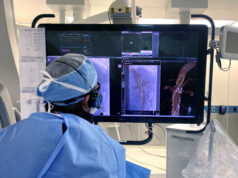
Based on the findings of their first-in-human feasibility study, Joost van Herwaarden (Utrecht University Medical Centre, Utrecht, The Netherlands) and colleagues write in the European Journal of Vascular and Endovascular Surgery (EJVES) that Philips’ Fiber Optic RealShape (FORS) technology “has the potential to improve intraoperative imaging”. Their results show that real-time navigation using FORS technology is safe and feasible in abdominal and peripheral endovascular procedures.
Van Herwaarden and colleagues detail that endovascular procedures are conventionally conducted using 2D fluoroscopy. However, a new technology platform, FORS, has recently been introduced, which allows “real-time, 3D visualisation of endovascular devices using fibreoptic technology,” they explain. This technology functions as an “add on” to conventional fluoroscopy and “may facilitate endovascular procedures”. The present first-in-human study assessed the feasibility of FORS in clinical practice.
The researchers performed a prospective cohort feasibility study between July and December 2018, recruiting patients undergoing either regular or complex endovascular aneurysm repair (EVAR) or endovascular peripheral lesion repair.
Van Herwaarden et al used FORS guidance exclusively during navigational tasks, such as target vessel catheterisation or crossing of stenotic lesions. In EJVES, they write that three types of FORS-enabled devices were available: a flexible guidewire, a Cobra-2 catheter, and a Berenstein catheter. They note that devices were chosen at the physician’s discretion and could comprise any combination of FORS and non-FORS devices.
The primary study endpoint was technical success of the navigational tasks using FORS-enabled devices, and secondary endpoints were user experience and fluoroscopy time.
The authors note that they enrolled 22 patients in the study, comprising 14 EVAR and eight endovascular peripheral lesion repair patients. They detail that, owing to a technical issue during start up, the FORS system could not be used in one EVAR. However, they report that the remaining 21 procedures went ahead without device- or technology-related complications and involved 66 navigational tasks.
Van Herwaarden and colleagues add that in 60 tasks (90.9%), technical success was achieved using at least one FORS-enabled device. In terms of user experience, operators graded FORS-based image guidance “better than standard guidance” in 16 of 21 and “equal to standard guidance” in five of 21 procedures. Finally, regarding fluoroscopy time, the authors report that this ranged from 0 to 52.2 minutes, adding that several tasks were completed without or with only minimal X-ray use.
The authors acknowledge some limitations of the present study. For example, they communicate that, although the operators had experience with the system in preclinical studies, they went through a learning curve during clinical use. Van Herwaarden et al write: “The optimal visualisation settings (regarding viewing angle, magnification, mono- or biplane mode, and optimal overlay) had to be identified for the different types of procedure.”
In addition, they note that workflow improvements, like the optimal positioning of the system, to work from both the groin and the arm, had to be learned during the study. “In this first study, the technical success would probably have been higher if more than two differently shaped catheters had been available,” they posit.
Van Herwaarden and colleagues list a few more limitations for clinical use, including the limited working length of the catheters and guidewire available for this study and also the inability to backload the guidewires. “These issues will be addressed in future releases of the system,” they state.
Finally, they relay that there were technical issues with the FORS equipment during this study. The technology could not be used in one patient, they detail, and using two FORS devices at the same time was impossible in on other patient. They note that these factors “probably affected study results”.
The authors conclude that this exploratory study “demonstrates the feasibility and potential of this technology in clinical practice and forms a foundation for future clinical research.” However, they recognise that comparative studies are needed to “prove and quantify” the benefits and potential radiation reduction for all types of endovascular procedures.


