A 53-year-old woman with a complex aortic malfunction causing a bulging blood vessel attended University Hospital Mainz (Mainz, Germany), having been rejected by several other hospitals due to the risky nature of her required surgery. Bernhard Dorweiler— head of the Department of Vascular Surgery at the hospital—utilised an emerging technology to assist in the careful planning of the surgery.
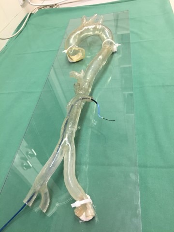
A Stratasys Polyjet (Stratasys Ltd) 3D printing process was used to convert MRIs of the affected site into an accurate model of the woman’s aortic arch. Rather than relying on what were ambiguous results from CT scans, Dorweiler and team found it much less complicated to use the 3D model to assess the extent of the patient’s problem.
“Looking through the CT scans, it was impossible to clearly visualise the anatomy,” says Dorweiler. “So we decided to 3D print a model, and it was then for the first time that it became clear what the origin and magnitude of the problem was.”
The surgery team from the University of Mainz Cardiothoracic and Vascular Surgery Department were not only able to visualise the problem, but could use the wholly unique model to explain the findings to the patient. Additionally, Dorweiler comments, “we even took it into each of the three surgeries as a point of reference during operation, which was crucial to the successful outcome.”
The hospital is a recognised Center of Excellence for cardiothoracic and vascular surgery, and regularly treats patients suffering from acute life-threatening aortic illness and problems requiring complex, patient-specific surgery. The adoption of additive manufacturing processes to assist in patient care is “crucial”.
“On average, CT scans with 1000–2000 images can be made per vascular-related patient case, which the surgeons use to analyse and diagnose the illness. This can be ambiguous and time-consuming when the issue is complex,” says Dorweiler. “With 3D printed models, we can quickly understand the individual patient anatomy and best determine the type of treatment required to successfully treat it.”
In the case of the 53-year-old patient, the surgical team were able to use the aortic arch to preoperatively simulate the planned procedure using a stent prototype to review accurate placement at the affected area. This process enables the surgeons to “practice surgery on the model repeatedly ensuring the correct design and fit of the stent implant the first time—significantly reducing time and cost in the operating theatre,” says a press release from Stratasys.
“As pointed out in current published studies, there are savings in operating time of 5–45 minutes when using 3D printed models prior to surgery,” comments Dorweiler. “Research is still ongoing, but if you take an average surgery time of 2–4 hours, you are looking at time savings of up to 40%. When you are dealing with complex vascular cases every day, these time-savings can be the difference between life and death.”
The technology also permits surgical trainees to rehearse endovascular procedures in accurate replicas of human anatomy, significantly contributing to their education.
“It is crucial that we continue to leverage the capabilities of 3D printing for medical training, education and research for future breakthrough-implementation,” adds Dorweiler.
“The pioneering use of 3D printing witnessed today underpins why the University Hospital Mainz is at the forefront of German medical research and development,” says Rene Martin, business manager of Healthcare EMEA at Stratasys.
“Leveraging high resolution 3D printing, the ability to replicate patient-specific anatomy is enabling physicians and surgeons to quickly plan, practice and determine life-saving surgical approaches – not only to improve patient care and outcomes, but also mitigate risk and reduce costs,” he states.
 clearance for aortic planning platform")


Thanks, how long and at what cost was the printing achieved?