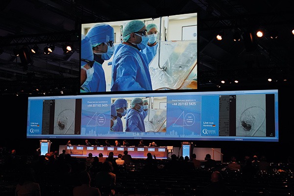
 Stroke is a major concern following TEVAR (thoracic endovascular aortic repair) and Charing Cross delegates heard the results from a collaborative study that pooled practice data from a number of high-volume centres in order to benefit patients undergoing the procedure. A highlight of the session was the transmission of a live complex aortic case from Hamburg, Germany, that illustrated the key learning points from the session. The case was a live endovascular arch reconstruction by Nikos Tsilimparis on a patient with thrombus on the arch.
The STEP (Stroke from Thoracic Endovascular Procedures) study was presented by the STEP investigators Roger Greenhalgh (London, UK), Stéphan Haulon (Le Plessis Robinson, France), Tilo Kölbel (Hamburg, Germany) and Fiona Rohlffs (Hamburg, Germany).
Advisors to the STEP study are Hugh Markus, a neurologist, Simon Redwood, an interventional cardiologist, Heinz Jakob, a cardiothoracic surgeon, and Kyriakos Lobotesis, a neuroradiologist.
“The purpose of this is to share the experience this group is producing. This experience includes over 800 procedures per year broken down into the various zones, and that data is a powerful tool. We also have the facts illustrated by a live procedure that is taking place alongside,” Greenhalgh said.
The STEP study aims to provide best practice for endovascular procedures for the ascending aorta, aortic arch, great vessel branches and high TEVAR to lower the risk of cerebral embolism. It is an independent, all-encompassing, open, interdisciplinary group built to learn from each other and to make TEVAR safer for patients. The STEP collaborators include Frank Arko, Carlos Bechara, Adam Beck, Dittmar Böckler, Matthew Eagleton, Dennis Gable, Stéphan Haulon, William Jordan, Tilo Kölbel, Gustavo Oderich, Jean Panneton, Geert Schurink, Santi Trimarchi, Marwan Youssef and the physicians who were all nominated by the manufacturers of the devices used in the procedure including Martin Czerny, Michael Dake, Ahmed Koshty and Rodney White.
The manufacturers of devices used by the 18 operators were Cook Medical; Gore; Bolton Medical, now Terumo Aortic; Jotec and Medtronic. The STEP study did not seek to compare outcomes based on the device used.
Rohlffs, noting that air, thrombus, particles or plaque can cause emboli during TEVAR, further explained that there was a STEP questionnaire sent out to gather data on experience and current practice. The questions were formulated before the data was collected. “With regard to the experience of the 18 physicians who were surveyed, all key opinion leaders operate on each zone and the survey group has a cumulative 143 years of experience in Zone 0 interventions; 193 years in Zone 1 and 232 years in Zone 2,” Rohlffs said.
STEP results
Illuminating data on the number of procedures performed within each zone per year revealed that there were 171 procedures performed in Zone 0 (21%); there were 135 procedures performed in Zone 1 (17%) and 510 procedures performed in Zone 2 (62%).
Other key findings include data from the preprocedural, intraprocedural and postprocedural phases of TEVAR.
Preprocedural results
Preprocedural findings showed there was 100% consensus that an interdisciplinary team is central to the final decision for treatment strategy in Zone 0. In Zone 1 and Zone 2, this was seen as less necessary.
The physicians unanimously responded “yes” to the question: Do you apply endovascular techniques in urgent or emergency cases? This established that the endovascular approach is undoubtedly utilised among those surveyed in this setting.
There was also 100% consensus to the question of which imaging technique was preferred to visualise the pathology, with all physicians revealing that they preferred CT angiography over MR angiography.
However, there was less consensus on how long before the procedure the imaging could be performed.
Intraprocedural data
With regard to intraprocedural data, there was consensus about anticoagulation and that the procedure should be done under antiplatelet therapy and that the activated clotting time should be above 250 seconds.
There was no consensus on revascularisation of the left subclavian artery, which was either done routinely or selectively and in terms of timing, sometimes in advance, simultaneously or on a case-by-case basis. There was 100% consensus on the use of cardiac output reduction in Zone 0, with less consensus in the other zones. There was no consensus on the technique of cardiac output reduction or embolisation prevention strategies with some physicians using carotid clamping, others using the CO2 flushing technique or minimising arch and device manipulation. There did not seem to be a role for filters here. There was no consensus on adjunctive techniques.
Post-procedural findings
All operators favoured CT angiography to visualise the stent graft at follow-up, but there was no consensus on the timing of the imaging. Again, there was consensus on antiplatelet therapy and postprocedural intensive care unit stay for the Zone 0 patients, but no consensus on the others.
There was no consensus on embolisation prevention techniques with some respondents using carotid artery clamping; others using a CO2 flushing technique and some respondents using other techniques, such as minimising archwire and device manipulation, and pacing.
Simon Redwood, King’s College London, spoke on the topic of reducing cerebral embolisation with transcatheter aortic valve implantation (TAVI) to note that stroke remains an issue and that it is under-reported. “The risk of stroke is up to 4–5% in trials at 30 days. Trials only report major disabling strokes, yet minor strokes or transient ischaemic attack have a two to four increase in mortality. Stroke risk is independent of operator experience and using diffusion-weighted MRI, 68–99% of patients show evidence of embolisation,” Redwood explained. With regard to the timing, stroke is a procedural issue and usually occurs within 72 hours of undergoing the procedure, he said.
Alan Lumsden (Houston, USA) presented useful data on transcranial Doppler being a useful technique for monitoring arch interventions. Transcranial Doppler is a non-invasive technique that uses a pulsed Doppler transducer for assessment of intracerebral blood flow. Cerebral emboli can be recognised by “high-intensity transient signals” (HITS). “Real-time middle cerebral artery monitoring is the gold standard as it relates immediately to intervention. It can be used for troubleshooting established and developing procedures and identifies high-risk manoeuvres,” he said.
Markus made the point that stroke is clinically relevant and it is what matters to the patient. It requires large numbers in studies as there are few endpoints. Surrogate endpoints such as brain imaging, imaging of emboli and cognition is often what counts, he noted.
Operative risk of stroke and death in endarterectomy studies is assessed differently depending on the specialty of the assessor, whether the assessor was the study author or whether it is a surgeon. This was one of the key messages that came out from Markus’ presentation that outcomes need to be assessed independently.
Markus further commented that transcranial Doppler cannot reliably distinguish solid from air emboli—and that the two types of emboli have very different consequences. “Transcranial Doppler emboli detection needs to be used with caution intra-operatively; post-procedure emboli are likely to be solid emboli. It provides additional information on cerebral blood flow,” said Markus.
Rohlffs had spoken earlier in the session on how systematic exclusion of air from unpacking to deployment avoids air embolism, explaining that the amount of air released by stent grafts can by minimised in several ways. Firstly, Rohlffs pointed to the way in which the amount of air released can be influenced by the ‘flushing technique’. Secondly, she observed that the construction of the stent graft system can influence the amount of air released during the procedure. Rohlffs maintained that synergistic effects of different approaches to reduce air release can be used, and concluded that “the amount of released air can be minimised.”
Haulon said that the STEP study has shed light on modern practice in the selected high-volume centres and that the group had learned from each other and were now ready to sit around the table to decide how they should perform these complex procedures. “We have learned about how we should design future studies and monitor results to find out what can be changed to reduce the stroke risk following complex TEVAR,” he said. Kölbel closed the session by saying this was an area in which there was a need for logical outcome measures, as very rough outcome measures had been used so far. “Additionally, it is vital to have independent measurement of outcomes as this has been shown to have a bearing on the data. There may also be some old dogmas to kill and I look forward to further collaboration,” he told delegates.
“We have worked out some of the questions we want to see answered and advances have been made in the areas of obvious consensus. The key next steps will be designing future studies that need to be carried out in order to benefit this group of patients who are inherently at risk of stroke after TEVAR,” Greenhlagh concluded.


