oowth
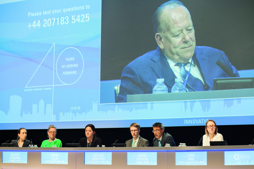
At the Charing Cross Symposium (CX; 24–27 April, London, UK) delegates heard compelling research-in-progress from a multidisciplinary team of surgeons, statisticians and health economists that the initial aortic diameter plus annual secondary sac diameter, as measured by ultrasound close to a patient’s home, could be a new way to follow EVAR patients. Prognostic modelling, based on data from the EVAR trials, was revealed to have predictive value in identifying patients at high-risk of secondary rupture and those who were classified as low-risk. This project was funded by the UK’s National Institute of Health Research Health Technology Assessment programme.
“Current EVAR 1 follow-up is suboptimal,” said Fiona Rohlffs, Imperial College, London, UK, who backed this statement using long-term data from the EVAR 1 trial, which was the first large randomised controlled trial conducted in the UK comparing open repair with endovascular repair of abdominal aortic aneurysms. The trial’s 15-year survival curve showed a benefit for EVAR patients in early follow-up, but this declined during the follow-up period of 15 years. Rohlffs called into question the surveillance schedule that was employed by emphasising data showing the proportion of EVAR patients returning for CT scan follow-up each year. “In EVAR 1, this was 90% in the first six months, but this declined to just 10% of the survivors by year nine,” Rohlffs explained.
Maarit Venarmo (Helsinki, Finland) then presented a contemporary EVAR dataset that showed exceedingly similar losses to follow-up in the surveillance protocol with time. The Helsinki dataset comprised 417 consecutive patients who underwent elective EVAR due to asymptomatic abdominal aortic aneurysm during 2000–2015. At six-months, there was follow-up data for 90% of patients. This declined to 40% at nine years, Venarmo said. The EVAR trials and the Helsinki University series were harmonised to achieve a comparable, consistent dataset.
Further, US data from follow-up in 20,000 Medicare beneficiaries between 2001–2008, published by Andres Schanzer et al in the Journal of Vascular Surgery, showed a 50% loss to imaging follow-up by year five.
Michael Sweeting, Cambridge University, UK commented that the multidisciplinary team wanted to develop a prognostic model for predicting future sequelae after EVAR. As background, Sweeting pointed out that the adherence to lifelong surveillance following EVAR was poor. “This is an ageing population; in the EVAR 1 trial, by six years, more than 40% of patients were aged over 80. Another reason why there might be poor follow-up is because there might be concerns about the frequent use of CT scanning (due to the radiation burden), and there is, therefore, a real need for patient-driven, safe and acceptable, truly personalised surveillance,” commented Sweeting. “Developing a prognostic model to be able to predict those who may be at high- and low-risk will help us to do that, so that we can potentially have less frequent follow-up, surveillance using different modalities (such as ultrasound) or potentially surveillance in primary care. There is interest growing in developing prognostic models to determine long-term outcomes following EVAR,” he explained.
The aims of this research, clarified Sweeting, are to model the trajectory of aneurysm sac diameters, to develop a prognostic model using model-predicted rates of sac growth and sac diameter in order to predict future events and to validate (in external data) its predictive accuracy.
The team developed the model using data from EVAR 1 and EVAR 2 randomised controlled trials for unruptured abdominal aortic aneurysms and validated the model using data from the Helsinki series.
Sweeting commented that prognostic models can be used to classify patients into low- and high-risk groups. “It is possible to reassess classification throughout follow-up using dynamic models. Predicted sac growth is important and outperforms observed change from baseline. It can be used to recommend reduced surveillance (for example biannually) or alternative surveillance (such as ultrasound).”
He reported on the diagnostic accuracy of the prognostic model with sac growth in predicting events over the next two-years to state that using the model, 40% of all patients could be classified as low risk and that it could also be used to correctly identify 85% patients who would go on to have a reintervention or rupture within the next two years. Roger Greenhalgh (London, UK), who was chairing the session, indicated that these were the key findings.
In an invited commentary, Stéphan Haulon, Le Plessis Robinson, France, endorsed the researchers’ approach regarding this data. “I fully agree that prognostic models will allow personalised follow-up and will probably provide better outcomes, if the patients that need follow-up actually get the follow-up. My concern is that after 15 to 20 years of treating aneurysms with endovascular procedures, we are still seeing a lot of secondary aneurysmal sac growth—is it because of technical issues? This is unlikely as we now have a 3D work station to properly design the endograft; access to the latest generation endografts and delivery systems that we can nicely position. However, despite this, we still have secondary aneurysmal sac growth and it is associated with an increased number of secondary procedures, an increased number of sac ruptures, and an increased inflammatory response,” he said.
Continuing, Haulon commented that EVAR is not succeeding (unlike open surgery) in treating aneurysms “because we are not resecting the sac, or closing the side branches and because we still have thrombus inside the aneurysm sac. We probably need to monitor the sac growth differently and maybe future endografts should include a way of easy monitoring, such as with a mobile phone that can actually be connected to the device to tell us about diameter pressure.
“I completely concur with your findings that with prognostic models we should leave the patients who will have an uneventful follow-up alone, and really focus on those patients who are high-risk, even though they are not a majority. It is very important information because if the patient needs to go to the hospital, wait a couple of hours, see somebody different, they will not come back. What you are suggesting is exactly what we should do.”
Greenhalgh then drew attention to “the key finding being the ability to classify 40% of all patients low risk” and its consequences.“And, further, the findings that 85% of those who will have a rupture in the next two years without investigation can now be identified. So this brings a whole different attitude to the follow-up of EVAR”.
He noted that certain assumptions have been made, including that ultrasound sac diameter will be available at, or close to home; that at the vascular centre clinical and CT angiography will be performed; that modern techniques will detect the cause of sac expansion and that rupture preventing reinterventions will correct sac growth.
Summarising these data Greenhalgh said, “it turned out that many patients did not accept having to go to hospital so often. The EVAR procedure requires lifelong annual surveillance. Lifelong surveillance has to be patient-friendly, simple and safe. From baseline morphology and annual sac diameter, low-risk patients can be identified and no hospital follow-up is needed. Further, high risk of secondary sac rupture in two years is predicted. The threshold for criteria of vascular centre referral needs further definition.”


