Spinal cord ischaemia may result as a consequence of both open and endovascular treatment of thoracic and thoracoabdominal aortic disease. Its clinical manifestations may range from numbness of the lower limbs to temporary or permanent paraparesis to complete flaccid paraplegia. It should be clear however that mobility impairment is only a part of the clinical syndrome that may result from spinal cord ischaemia. Incontinence, bed sores, and recurrent infections may cause terrible problems in the daily life of the patients and are psychologically very poorly tolerated often resulting in severe depression, writes Germano Melissano.
Unlike patients with post-traumatic paraplegia, postoperative spinal cord ischaemia often affects elderly subjects with significant comorbidities. This explains the very poor survival rate of subjects with the more severe forms of the condition. Moreover, only a fraction of these patients return to a socially productive life while most of them require on-going assistance at specialised institutions.
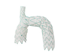
The pathophysiology of spinal cord ischaemia is complex, multifactorial and still poorly understood, while the mechanisms involved are different in open and endovascular procedures. When feasible, endovascular treatment of the thoracic and thoracoabdominal aortic disease (thoracic endovascular aortic repair—TEVAR) reduces the morbidity of surgical access and eliminates the need for aortic cross-clamping. However, comparable rates of ischaemia are reported with both approaches.
While the key factors for prevention of spinal cord ischaemia during open and endovascular procedures are different, they are based on the same following basic strategies:
1) Minimise spinal cord ischaemia time
This may be achieved in open surgery by reducing aortic clamping times, by sequential aortic clamping and by using distal aortic perfusion by means of circulatory assistance. In endovascular procedures it may be important to revascularise the left subclavian artery before TEVAR and to obtain pelvic and lower limb reperfusion as early as possible.
2) Preserve spinal cord blood supply
Nowadays it is often possible to directly visualise the spinal cord vasculature with preoperative diagnostic tools such as angio-computed tomography (CT) and magnetic resonance imaging. This may allow selective re-implantation of critical intercostal arteries during open surgery and limitation of aortic coverage by stent-graft, avoiding unnecessary sacrifice of intercostal arteries.
In both open and endovascular procedures it is important to preserve subclavian and hypogastric contribution as well as to stage procedures, when possible, in order to avoid simultaneous interruption of several different districts of spinal cord feeders (ie. subclavian, thoracic, lumbar, hypogastric).
3) Increase tolerance to ischaemia
While hypothermia may be employed in open surgery to increase tolerance to ischaemia of the spinal cord, pharmacologic neuroprotection—which has not been very effective so far—may be a good field of research for future applications for both open and endovascular procedures.
4) Optimisation of spinal cord perfusion
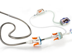
Optimising spinal cord perfusion by raising arterial systemic blood pressure and reducing cerebrospinal fluid pressure are both very important. It should be noted that cerebrospinal fluid drainage is not free of risks so this should probably be offered selectively to patients at greater risk for spinal cord ischaemia. Moreover, an automated apparatus for cerebrospinal fluid pressure and drainage monitoring may reduce the incidence of complications.
Haemodynamic stability is absolutely crucial and so is intra- and postoperative control of haemorrhage and transfusion requirements. Equally, an intensive care team that is familiar with all aspects of postoperative care is invaluable in maintaining haemodynamic stability in the postoperative period.
In order to avoid haemodynamic instability, which is secondary to perioperative cardiac events, we advocate the use of coronary CT scans in all patients undergoing elective thoracoabdominal repair and coronary angiography in patients with positive CT findings. A significant number of those patients require coronary angioplasty with non-drug eluting stents and the aortic procedure is postponed for one month under single antiplatelet therapy.
5) Early detection of spinal cord ischaemia
It has been shown very clearly that spinal cord ischaemia may respond positively to treatment with clinical improvement or even complete resolution, provided it is diagnosed early enough, ie. before ischaemia has evolved to infarction and permanent paraplegia. Neurologic monitoring of cord function can be performed in anaesthetised patients with somatosensory evoked potentials (SSEP) and motor evoked potentials (MEP) or both. The intraoperative detection of spinal cord ischaemia using MEP and SSEP monitoring gives a surgical team the opportunity to promptly trigger interventions for maximising spinal cord perfusion and to potentially reverse spinal injury. Postoperatively it is important to assess neurological status as early as possible.
While no single method has been able to eliminate or even significantly reduce the incidence of spinal cord ischaemia during thoracic aortic procedures, a multimodal approach may be pursued that wishes to:
- Improve the preoperative fitness of the patient;
- Assess spinal cord anatomy using advanced imaging techniques;
- Tailor an individualised open or endovascular strategy to treat the aortic pathology;
- Ameliorate the intraoperative surgical and anaesthesiologic management of patients;
- Reduce postoperative complications.
The synergic effect of improvements in many different areas offers the best hope of further reducing the incidence of spinal cord ischaemia in the future.
Germano Melissano is an associate professor of Vascular Surgery at the Università
Vita-Salute San Raffaele, Milan, Italy


