In Tuesday’s CX Congenital Vascular Malformations session, delegates heard about how approaching congenital vascular malformations with systematic pathways of care and classification systems can greatly assist physicians in achieving consistently successful positive outcomes.
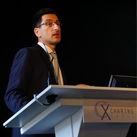
Joe Brookes (London, UK) presented on classification of arteriovenous malformations, charting its “emerging clarity” in recent years, and noting that these advances hold a great deal of promise for the future treatment of these conditions.
“Although extracranial high-flow arteriovenous malformations (AVM) have been recognised since the days of the great Middlesex Hospital surgeon John Bell, the understanding of this complex group of disorders has continued to elude us until recently,” Brookes told delegates.
AVMs are “mercifully rare”, Brookes said, comprising only 10–20% of all congenital vascular malformations, of which only 5% are extra-cranial. They are understood to be early (4–6 week) embryological failures of vascular germ line cellular differentiation (angiogenesis). This is in distinction to vascular tumours which mostly represent a post-natal acquired loss of cellular differentiation. Although there are some rare genetic variants (eg. hereditary haemorrhagic telangiectasia; an autosomal dominant familial disorder), most high flow AVMs remain genetically elusive and of apparently random aetiology.
“Successive attempts to provide an encompassing method of classification of high-flow AVM have been proposed since the early 1980s and spearheaded since 1992 by the International Society for the Study of Vascular Anomalies (ISSVA), most recently meeting in Melbourne, Australia, in April 2014 and due to meet again 2018,” Brookes explained.
Brookes believes, “Most exciting has been the recent recognition of the therapeutic approach to AVMs developed and championed by Wayne Yakes (Denver, USA) leading to publication in 2014 of the ‘Yakes classification’,” which provided a “relatively simple” pattern-based recognition method of classification correlating to specific curative treatment methodologies, seeking to achieve endothelial ablation of the venous side of the AVM nidus.
These methods, Brookes told the audience, are “reproducible in daily practice such that there is a degree of emerging clarity in this erstwhile confusing field and thus some genuine hope at last for our patients who suffer with these distressing conditions.”
In his presentation, Andreas Saleh (Munich, Germany) laid out a set of vascular anatomy imaging principles to follow. First, patient history should be analysed, followed by clinical examination and colour coded duplex sonography (CCDS). Whether the anomaly is high- or low-flow should then be established with T2-weighted imaging. CCDS or contrast-enhanced magnetic resonance imaging can be used to distinguish low-flow malformations, while fat-suppressed T2 images in three planes can be used to assess the extent of a slow-flow malformation, Saleh noted.
Saleh cautioned that contrast should not be used when the type of slow-flow malformation is used, and angiograms should not be performed in extratrucular slow-flow malformations. Making a distinction between microcystic and macrocystic lymphatic malformations is important, and operators should avoid confusion by haemorrhage, phleboliths and thrombi. What parts of the malformation are vascular and non-vascular needs to be determined, and care should be taken that hypervascularity of infection or tumour is not consued with AVM. Once this is assured, Saleh said, cross-sectional imaging will be used to locate the nidus and the draining veins.


