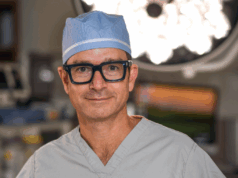
Due to its two-dimensional (2D) nature and the high radiation exposure associated with its use, fluoroscopy is an imperfect solution to endovascular surgery’s need for intraoperative imaging. At the 2016 VEITHsymposium (15–19 November, New York, USA), Matthew Eagleton, Cleveland Clinic, Cleveland, USA, presented his centre’s initial experiences with the Intraoperative Positioning System (IOPS; Centerline Biomedical), suggesting that it may represent an opportunity to transition away from fluoroscopy.
Eagleton told VEITHsymposium delegates that the IOPS system provides interactive enhanced 3D vascular images, as well as “electromagnetic tracking of endovascular tools within the vascular tree, correlating directly with in vivo localisation”. With the ability to manipulate the images shown, the system also allows operators multiple and ideal views. These imaging and navigation capabilities, Eagleton said, are “superior to standard 2D fluoroscopy”.
The system is based on a mathematical model for vascular image construction from DICOM CT imaging data, and the model was then tested by assessing the relative geometry of the aortic branches. Though the testing was aortic, Eagleton noted that the system “is not limited to the aorta”. Eagleton also explained that the algorithm used is automatic and patient-specific, with an image of the aorta generated using a DICOM-DICOM overlay. This overlay can then be used to create an overlay fusion of the mathematical model.
Once the model was created, Eagleton and colleagues needed to be able to see where on the model they were moving without using fluoroscopy, so began to work on device localisation methods. A navigation system was developed using sensors on sheaths, wire and catheters, in combination with patient sensors, used to overcome patient and table movement. Display systems were developed to allow Eagleton and colleagues to view images in parallel to angiography imaging based on fluoroscopy. Bench-top testing with several aortic models was performed before transitioning to the operating room. Eagleton noted, “The biggest challenge in this transition was overcoming the electromagnetic interference from the C-arm, but we were able to do it.”
A pre-clinical study in a porcine model was conducted, running the IOPS in parallel with standard fluoroscopy. The initial goal was to assess the system’s ability to cannulate visceral vessels, which would be verified with angiography. Showing examples of the imaging, Eagleton reported, “One of the benefits of this was that we were able to re-orient the 3D image to provide optimal views. We were also able to magnify the images without any additional use of radiation.”
Since the pre-clinical test, the system has been transitioned to clinical application and used during the placement of branched endografts for two patients.
Following the initial encouraging experience, Eagleton suggested that there could be refinement of sensored endovascular tools, including catheters, wires and sheaths. The sensitivity and specificity testing could also be improved, he continued, compared to the gold standard of live fluoroscopy, while the enhancement of vessel deformation predicting should also be explored.
“Non-fluoroscopic imaging and navigation will replace all of our current modalities for endovascular intervention,” Eagleton concluded. “We must strive for improved procedural imaging, improved procedural navigation and limited radiation exposure.”


