In a session on “Reduction of radiation challenges” at CX Aortic Vienna 2021 (5–7 October, broadcast), experts highlighted new technologies with the potential to make aortic surgical procedures simpler and shorter, thereby reducing radiation exposure to patients and care teams. A presentation by Tom Carrell (Barrington, UK) on intelligent maps for surgery and an edited case by Tilo Kölbel (Hamburg, Germany) were among the programme highlights to emphasise this key message, while a Philips-sponsored satellite symposium saw early users of Fiber Optic RealShape (FORS) speak about their key experiences with this developing technology.
All sessions are available to view on demand. Click here to register and access the recordings.
Intelligent maps for surgery offer increased accuracy and reduced radiation
Opening the session, Tom Carrell (Barrington, UK) outlined the benefits of Cydar EV Maps, which recently received regulatory clearance in the European Union. This software includes integrated planning, navigation and review, offering a dynamic, three-dimensional (3D) map of the current state of a patient’s anatomy, he informed viewers. Carrell relayed that the technology offers more accurate device positioning and better overall outcomes with a reduced radiation dose, and that each new patient’s plan and surgery is informed by all previous similar cases.
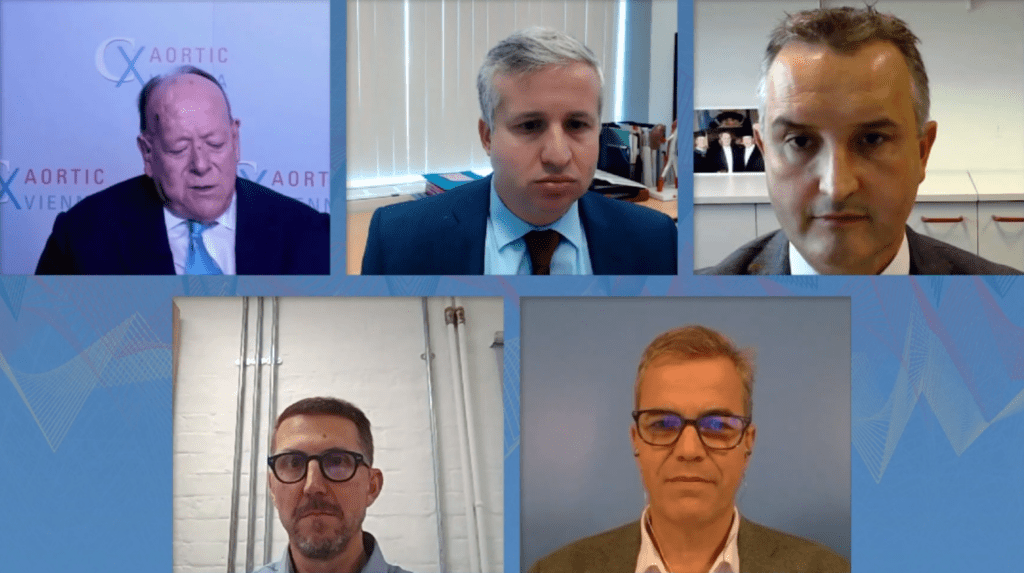
Moderator Andres Schanzer (Worcester, USA) congratulated Carrell on an “excellent and provocative talk”, going on to ask about the applicability of Cydar EV Maps. Carrell responded by stressing that Cydar EV Maps is a software and is therefore generally applicable.
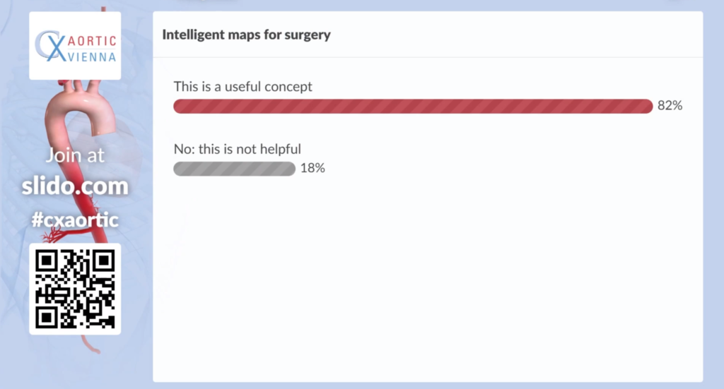
Audience polling following Carrell’s talk revealed that 82% of voters in the audience believe intelligent maps for surgery are a useful concept.

Registrants can view Carrell’s presentation on demand here.
FORS provides “new, precise and low-radiation visualisation tool”
In an edited case, Tilo Kölbel (Hamburg, Germany) presented a branched endovascular repair for a type IV thoracoabdominal aortic aneurysm using FORS laser light guidance. He concluded that the procedure is feasible, with a low radiation dose and short fluoroscopy time, and that the 3D visualisation FORS offers enhances target vessel catheterisation. Kölbel summarised that FORS provides a “new, precise and low-radiation visualisation tool for complex endovascular aortic repair”.
Moderator Joost van Herwaarden (Utrecht, The Netherlands) was keen to know what the learning curve is for this novel technology, with Kölbel responding that FORS is “very intuitive”. An audience question addressed potential fatigue issues with fibre optic cables, but Kölbel stressed that, in his experience, the cables “usually last the whole case”.
Reflecting on Carrell’s presentation and Kölbel’s case, anchor Roger Greenhalgh remarked: “We have shown the audience the future”.
Registrants can view Kölbel’s edited case on demand here.
Early users share key moments with FORS 3D device guidance
In a Philips-sponsored satellite symposium, Kölbel opened a discussion on early users’ key moments using FORS 3D device guidance. Schanzer, Geert Schurink (Maastricht, The Netherlands), van Herwardeen and Marc Schermerhorn (Boston, USA) all shared their experiences. Throughout the session, audience members were also encouraged to highlight what they believe to be the key benefits of FORS technology by contributing to a word cloud. The benefit of 3D visualisation was deemed to be the most important element, closely followed by precision and reduced radiation.
Click here to watch Philips’ satellite symposium (no paid registration required).
Also in the “Reduction of radiation challenges” session, Michele Antonello (Padua, Italy) spoke about an off-the-shelf endograft with four inner branches to treat thoracic aortic aneurysm, which, used in conjunction with intravascular ultrasound (IVUS), has resulted in shorter procedural times in his experience.
Registrants can view Antonello’s presentation on demand here.
Finally, Alun Lumsden (Houston, USA) argued that dynamic computed tomography (CT) and magnetic resonance (MR) represent the “gold standard” for aortic imaging.
Registrants can view Lumsden’s presentation on demand here.


