 The management of type B aortic dissections represents a significant challenge for the vascular community. Despite the great advances obtained since the development of endovascular treatment, questions still remain on how to best treat complicated and uncomplicated dissections, and when. Trying to seek consensus on how to address the different scenarios, Cook Medical held its second Global Dissection Forum, a gathering of experts who discussed the most pressing topics surrounding dissection-specific devices, the aspects involved in the decision of whether to intervene or not, the time of intervention, and techniques for false lumen thrombosis.
The event, which took place at the recent Charing Cross International Symposium (25–28 April 2017, London, UK) was moderated by Eric Verhoeven, Nuremberg, Germany.
In his introduction, Verhoeven stressed the importance of the subacute phase in the classification of type B dissections and highlighted the vascular community’s interest in a paper published by his group last year in the Journal of Cardiovascular Surgery, which discussed treatment algorithms for patients in the (sub)acute phase. As with the ideal time for intervention, the jury is still out for other topics related to type B dissection. Verhoeven commented: “For complicated type B dissections, we all agree that the choice is TEVAR plus adjuncts; however, we still have questions: do we use the PETTICOAT technique, or do we go for the STABILISE technique? But also, when? What are the limitations? When should TEVAR be avoided?”
In recent years another point of intense discussion has been the management of uncomplicated type B dissections. “Should we stent immediately, with hyperacute TEVAR? Or should we wait a few weeks and then stent it (2–12 weeks)? Or should we stick with best medical treatment, as a number of people still believe we should? Best medical treatment alone is not ideal, and we now know from IRAD about late complications,” Verhoeven said.

In addition, he noted, the INSTEAD XL trial “surprised us” with results showing best medical treatment alone yields worse outcomes than TEVAR in uncomplicated type B dissections. This was also shown in the ADSORB trial, but with smaller numbers. So the big questions, according to Verhoeven, are: Should we treat uncomplicated type B dissection? How can we treat these patients without serious complications to justify treatment and avoid late complications? What subgroups need treatment, and what is the optimum time of treatment?
Regarding time of treatment, Verhoeven referred to a paper by Matt Thompson’s group showing that “in the acute phase you have some complications with TEVAR, but in the subacute phase you do not. Patients who do not need the hyperacute treatment should maybe wait. The dissection flap remains flexible in the subacute phase like in the acute phase.”
Verhoeven went through some of burning questions in the treatment of chronic dissections and post-dissection aneurysms with fenestrated/branched grafts: “Do we need to increase graft oversizing? Do we need to extend the coverage of the aorta? Do we need to walk in the direction of inner branches? Certainly we need to place longer bridging stent grafts to have better sealing in dissected vessels.”
Minimising risk
In the following presentation, Bijan Modarai, London, UK, spoke about how to minimise procedural risk when managing type B dissections. Every type B dissection patient is different, he said, and physicians “have challenging decisions to make about timing of intervention, how to intervene, and how to intervene safely”. One of the most effective ways to minimise the risks associated with intervention is to wait, “get out of the hyperacute phase, perhaps with intense imaging to make sure that intervention is the right approach for the patient”.

When the decision to intervene is made, one of the most important factors to consider is the proximal extent of the repair, Modarai noted, and added, “We know from the literature that almost half of all repairs will be in zone 2; dissection patients are younger than those with aneurysmal disease and tend to have less favourable arch morphology; and our data suggest that around 16% of cases will have a retrograde extension into the arch. According to the IRAD data, the latter does not impact adverse outcomes but all of these factors are important considerations for making the decision whether to intervene or not and how safe that intervention is likely to be, particularly in ‘uncomplicated’ cases.”
Unfavourable arch anatomy is associated with increased risk. It demands a longer sealing zone, and tortuosity and high angulation make deployment more challenging. “The characteristics of the graft and the reliability with which we deploy it are of paramount importance. Particular attention should be paid to selecting a healthy and lengthy seal zone which may require extraanatomical debranching of the supra-aortic trunks, even in the acute patient. You can obtain good conformability, precise deployment and a much better result. Also, going closer to zone 2 has its own risks—we know there is higher rate of type A retrograde dissection,” he said.
So what have we learnt on risk factors associated with retrograde dissection? Modarai commented that “stents are rigid and this causes a compliance mismatch”. Oversizing is also used in the acute scenario but excessive radial force has its problems. The jury is still out on bare stents. There are some data to suggest that if you have an enlarged ascending aorta that may put the patient to extra risk.
Should these patients have transoesophageal echocardiography as a completion check? “We should have a low threshold for doing investigations like this, particularly in patients who we suspect are at risk of a retrograde dissection,” he said. In terms of oversizing, a study by Liu et al (J Endovasc Therapy 2016) of acute and chronic type B dissections and employing an average oversizing of 10%, showed a significantly higher rate of retrograde type A tears in patients whose grafts had been oversized >5%. With the oversizing of less than 5% there were no cases of retrograde type A dissection. “Perhaps we should not be doing any oversizing but the effect of this strategy on proximal seal is unknown,” Modarai told the forum.
He commented “we are still in the infancy of predicting what happens to the aorta when it is stented. We will soon be moving to a system in which we use algorithms to predict the patients that will have problems and how the stent graft will impact haemodynamics within the aorta. Currently we size our stents based on 2D axial images that do not take into account the cardiac cycle and 3D structure. This type of technology will help us in the near future. Additionally, when your computer programme tells you how much to stent within the aorta it will take into account the risks associated with where you end the distal extent of your repair.”
Moving on to the topic of spinal ischaemia, Modarai noted that the rate of this complication is low in dissection but “there is a price to pay for covering more aorta and there is particularly risk the more distally you cover. We also need to know more about the management of the collateral circulation in the acute scenario. If you cover the left subclavian we have some indicators whether we ought to revascularise or not. But actually in the acute scenario we usually cover the left subclavian and deal with the consequences later on.”
On the subject of monitoring signals of spinal cord ischaemia, the “new kid on the block” is near-infrared spectroscopy, Modarai said. The system has pads that are placed in the thoracic and lumbar region and monitor the paraspinal collateral circulation. One of the important aspects is that anaesthetics do not affect the readings. “Hopefully soon we are going to have many devices that are tailored for dissection, and we need to keep an open mind for novel adjuncts,” he concluded.
In a discussion following Modarai’s presentation, Stéphan Haulon, Lille, France, questioned what the ideal device design would be in order to achieve a proximal sealing zone in the arch, maintain flow to the left subclavian artery and completely exclude the dissection. He added, “How far would you go into the arch, routinely, for acute type B dissection? You probably would need to go all the way to the left common carotid artery with the graft. And in the middle of the night the patient has a life-threatening condition and you decide to cover the left subclavian artery, using a short graft, as you just want to restore flow into the true lumen so the patient might survive. You also do not always completely occlude the proximal entry tear because it actually goes inside the left subclavian artery origin so you would need to put an occluder in it. So if there was a graft with a proximal scallop for the left common carotid and retrograde branch for the left subclavian artery, would you use that routinely if you had it on the shelf?” Modarai said that if there was an off-the-shelf solution he would use it; however, he added he would be “slightly concerned about the extra manipulation in that type of fragile aorta.”
Verhoeven then, on another topic, asked if enough attention was being paid to the positioning of the proximal stent or stent graft in such a way that it is lies perpendicularly to the wall and distributing the radial forces: “We heard the advice not to oversize and do a bit less, but both proximally and distally it is extremely important to make sure that this far more flexible graft is lying very perpendicular to the wall and the forces are distributed.”
PETTICOAT concept
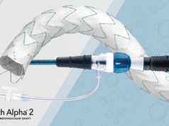
Joseph Lombardi, Camden, USA, focussed on the PETTICOAT technique for acute type B dissection. The technique was used in both STABLE I and STABLE II studies—these studies employed a unique composite thoracic endovascular aneurysm repair construct (proximal stent graft and distal bare metal stent—Zenith Dissection, Cook Medical) for the treatment of complicated type B dissections.
STABLE I had broad inclusion criteria, with an earlier iteration of the device. The current model, with a nitinol base dissection stent, was the one used in STABLE II. In this device the stent graft (TX2) has had all the barbs removed and is fixated exclusively by radial force, in addition to the nitinol bare metal stent distally, which supports the true lumen and is placed across the visceral vessels. It is also available in tapered shapes. The device has been designed specifically for dissections.
STABLE I enrolled 86 patients; the primary endpoint was 30-day mortality and had follow-up through five years. In terms of mortality within 30 days, for acute dissections it was 5.5% and for non-acute it was 3.2% (overall rate 4.7%). “In STABLE II we looked at patients with rupture or malperfusion, so we narrowed the inclusion criteria to the most serious of complicated varieties of dissection,” Lombardi said.

The survival rate at 30 days was similar for STABLE II and STABLE I and comparable to the SVS dataset. He explained that the stroke rate in STABLE II was similar to that reported in the SVS dataset and other registries. “Stroke is the biggest bad actor that we have in the acute setting of dissection, and it has a lot to do with wire and catheter manipulation of the aortic arch. We discussed revascularising the left subclavian artery as a means for stroke prevention—is it worth it in the acute setting, where most strokes are bihemispheric, anterior and posterior? In the acute setting we have a very fragile aorta that is coated with platelets and thrombogenic material that likely the source of these strokes,” he noted.
In terms of paraplegia, the rate in STABLE I was 1.2%, Lombardi commented, “but the strategy with that study is that we only needed to cover the entry tear, so we did not need to extend down with a covered endograft. We supported the remainder of the true lumen with a dissection stent. Based on that, we had an incredibly low rate of paraplegia. Then you look at STABLE II, in which we have a 5.5% paraplegia rate. In this trial we did extend down the aorta more frequently. Our investigators wanted to cover more, and more coverage of the thoracic aorta meant more false lumen thrombosis. So our paraplegia rates went up.”
Lombardi told participants that any application of TEVAR for type B dissection should not be seen as the full treatment “but as a management tool”, and added “when you do a TEVAR for dissection, it is your initial stage of management. If you try to win the game with that one procedure you are going to have a lot more complications than you could have had.”
What are the benefits of the PETTICOAT technique? “We have shown that mortality and paraplegia rates are low, and that the accumulative all-cause mortality in STABLE I at five years is 16%. That was impressive. The bare stent has utility later on in the 30–40% of patients who experience aortic growth after TEVAR. Having that dissection stent there gives you options. It allows you to come back if there is growth and reintervene in the torn vessel without the use of any other aortic stent grafts, just a small covered stent at the viscerals. You could shut down that re-entry flow through the visceral vessel and ultimately induce false lumen thrombosis. All you need is a covered stent and some interventional skills,” Lombardi said, and added, “Overall, this technique gets people out to trouble to live and fight another day. There is still a risk of disease progression that requires close surveillance and reintervention. The bare stent management can be advantageous in the early and long term with the understanding that management of complicated type B dissections is a long-term commitment.”
Verhoeven commented that most people do not realise that STABLE II had very challenging type B dissection cases, and questioned if the dissection stent should be used also in moderately complicated patients (without rupture or malperfusion)?
Lombardi commented: “Our investigators did not use dissection stents in all cases. In those that were for focal dissection or exclusive to the thoracic aorta, you probably would not need it. However, for extensive dissections I would use the dissection stent because early remodelling is very favourable and it would give me options later on if needed.”
Stabilise or obliterate?
Jean-Marc Alsac, Paris, France, spoke about STABILISE (Stent-Assisted Balloon- Induced Intimal Disruption and Relamination in Aortic Dissection Repair), a technique to obliterate the false lumen in type B dissections. He referred to a paper from IRAD in 2013 showing the occurrence of aortic growth after TEVAR in two thirds of patients at five years, and said that that was seen also in the INSTEAD XL trial. “Closure of proximal tears is efficient but not always sufficient in the long term—we still had 27% of patients in the TEVAR group with aneurysmal progression at five years,” he noted.
Adding a bare stent (as in the PETTICOAT technique) is useful, especially in acute patients with dynamic malperfusion, Alsac said. In a paper published in 2014, his group described an experience with 15 consecutive patients with acute dynamic malperfusion. All were treated with PETTICOAT with good results and all recovered well. He commented: “In the acute phase, it gave us very good results but the problem is that in the long term we faced the same incomplete remodelling results. Abdominal re-entry tears still lead to thoracoabdominal growth”.

With the STABILISE concept the idea is to induce intimal disruption by ballooning the bare stent in order to relaminate the whole aorta. It needs a number of balloon expansions to provide relamination of the aorta. Alsac’s group has three years of experience with this technique. Of 121 TEVAR procedures for acute type B dissection patients, 52 were treated with STABILISE. In a mean follow-up of 13 months, there were no late adverse events (aortic rupture, stent migration, intimal flap erosion, redissection). He told participants that the STABILISE approach is “a feasible endovascular technique that shows promise to achieve complete repair of the dissected aorta by inducing complete false lumen obliteration. The restoration of uniluminal flow in the thoracoabdominal aorta has the potential to improve long-term outcomes. Prospective, multicentre investigations are required to implement this strategy more broadly, and to define the best indications and timing for such an aggressive therapeutic option.”
In conclusion, Alsac said, long-term results of TEVAR are disappointing. “Patients experience aneurysmal growth and need strict surveillance and restrictive medical treatment. Should we have a more aggressive aortic repair in the acute phase of selected patients? In our experience the STABILISE technique is safe and reproducible. It is efficient to treat acute complications and seems to prevent aneurysmal progression. It has a low reintervention rate and decreased overall morta-l ity during follow-up. It has become our first line technique for complicated acute, subacute, chronic, and Marfan aortic dissections,” he said.
Candy-plug

Another technique for false lumen occlusion, the candyplug approach, was described by Tilo Kölbel, Hamburg, Germany. Kölbel noted that in chronic type B dissection, false lumen perfusion prevents aortic remodelling and leads to late death in one third of the patients. The reason for this is probably that in the false lumen, perfusion and pressure keep unchanged due to the presence of intercostals and bronchial arteries originating from the false lumen.
“What we should do besides extending our coverage down to the coeliac artery is occlude the false lumen with embolisation techniques using plugs, coils, glue, candy-plug or the knickerbocker technique. This does not restrict further distal techniques like fenestrated EVAR,” Kölbel said.
Iliac occluders are one of the available tools but “their maximum diameter is 24mm”, he added. For this reason, and because the false lumen is often much larger in diameter, Kölbel’s group, in collaboration with Cook Medical, developed a larger plug (50mm) that we combine with an iliac occluder. “If you do this, even if an aneurysm has been growing after TEVAR, you occlude the false lumen, the entrance from below, the aneurysm tends to go smaller and the candy-plug and the true lumen stent graft adapt to each other. In Hamburg, between 2013 and 2016, 18 cases were performed. There were no procedural complications, no spinal cord ischaemia, no deaths at 30 days, and no aneurysm expansion at six months.
Chronic dissections
Stéphan Haulon then spoke about chronic dissections and patients with prior repair for type A dissection and enlargement of the arch and descending aorta. Challenges associated with the treatment of this subgroup of patients include a difficult distal sealing zone, working in a narrow true lumen, the fact that target vessels are perfused by the false lumen and limited experience.

Haulon said that when the enlargement extends distally a fenestrated distal extension is usually the solution. He showed an example of a 50-year-old patient who in 2009 underwent ascending aortic replacement for acute type A dissection, followed by a redo sternotomy in 2013, including arch repair and elephant trunk. The patient then underwent TEVAR from the elephant trunk to the coeliac trunk and then an aorto bi-iliac open repair with the objective to perfuse both internal iliac and distal lumbar arteries (to reduce the spinal cord ischaemia risk), and the final step was the placement of a fenestrated endograft between the TEVAR and the distal open repair graft. The patient had a tear at the level of the right renal artery, which was bridged with a stent. CT scans after the TEVAR procedure and then after the fenestrated graft showed complete occlusion of the false lumen and the expansion of the true lumen after the placement of the fenestrated device.
He moved on to show a second case in which the right renal artery originated in the false lumen, with entry tears situated above and below the artery. The patient was treated with a staged procedure—the first stage consisted of left subclavian artery branched TEVAR, and the second stage consisted of fenestrated-branched EVAR.
He noted that “it should not be a competition between endovascular and open procedure and at every stage you have to figure out what is the best solution for the patient”.
This article was supported by Cook Medical.


