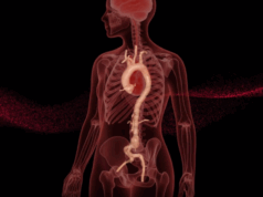
In the Aortic session at this year’s Controversies and Updates in Vascular Surgery meeting (CACVS; 23–25 January, Paris, France), Stéphan Haulon and Dominique Fabre will discuss the rise of artificial intelligence (AI), informing delegates how this technology could be the solution for aortic aneurysm follow-up. Here, they give a summary of their key points.
AI applied to medicine has been growing exponentially in recent years, according to the number of scientific publications in the field. Its efficiency at characterising and learning from large amounts of data makes it well suited to tackle the challenges of medical imaging. And challenges, there are many: ageing population, systematic use of imaging at every step, new generation equipment with a 40-fold increase of output images over the last 30 years.
Overwhelmed radiologists are asked to read thousands of images daily while everything suggests that a very needed revolution is coming: a large volume of digital data, unprecedented computing powers and detection or characterisation questions that are often well defined. The training of high-performance machine learning models has become a reality.
Aortic aneurysms diameter measurements fit as an ideal candidate to benefit from more data-driven automatisation: it is a tedious and time-consuming task with high inter- and intra-operator variability.1 While there are several commercially available tools to assist on these measurements, none provide full automatisation. Most are efficient in segmenting the lumen of the aorta but often fail at capturing the full diameter of the thrombus (less contrasted on CT scans). Aneurysm measurement is performed to decide whether to intervene and during follow-up after open and endovascular repairs.
To address this challenge, we initiated a partnership with a young startup specialised in AI in Medical Imaging (Incepto Medical, Paris, France) using Machine-Learning-based solutions. In this project, we first retrospectively collected a large number of CT scan examinations from patients who benefited from an abdominal or thoracic aortic aneurysm endovascular repair at Marie Lannelongue Hospital, France. All pre- and postoperative images available were included, providing a large variety of cases, with various segments of the aorta, various lesion sizes, in the presence of endografts or not, evolving or not, and from different hospitals and therefore CT manufacturers and models. On all of these examinations, hundreds of contrasted CT scans were segmented manually to constitute a large segmented aorta database with their outer maximum diameters. We then built a pipeline of algorithms. A first one, on the localisation of the aorta area in chest and / or abdominal CT scan. A second one, on the segmentation of the localised aorta. These two algorithms are 3D Fully Convolutional Networks models. A third one, on the computation of the central line of the segmented aorta. And finally a last one, on the measurement of the maximum external diameter perpendicular to the central line, the smallest axis.
The results, although preliminary, show a good performance in segmentation of the aorta by including the outer wall of healthy zones and dilated zones with the thrombus. The results are also quite high in cases with the presence of stents.
Usual next steps of this work will include an increase of the training database, by manually segmenting more aortas, internal and finally external validations. For internal test set, two experienced operators will manually measure on a large variety of cases (with and without endograft, on normal and aneurysmal aortas), external aortic diameters at 8 levels of the vessel. This ground truth will be used to assess the algorithmic pipeline performance, inter and intra-reader variability.
Several recent works have been published around automatic aortis segmentation. But to our knowledge, our database of hundreds of exams is by far the largest2–4 and the only one to address the entire aorta, from the ascending aorta down to the iliac arteries, when others have only addressed the abdominal portion of the aorta. Another originality of our approach was to consider a large amount of exams (80% of our database) where a graft was present in the aorta, against only 6 cases out of 40 in Lareyre et al and none in Lu et al. Finally, the close collaboration between surgeons and data scientists allowed us to elaborate a tool specifically designed to fit smoothly into the clinical workflow.
References
- Mora et al. Eur J Vasc Endovasc Surg. 2014
- Lu, Jen-Tang, et al. “DeepAAA: Clinically applicable and generalizable detection of abdominal aortic aneurysm using deep learning.” International Conference on Medical Image Computing and Computer-Assisted Intervention. Springer, Cham, 2019.
- López-Linares, Karen, et al. “Fully automatic detection and segmentation of abdominal aortic thrombus in post-operative CTA images using Deep Convolutional Neural Networks.” Medical image analysis 46 (2018): 202–214.
- Lareyre, Fabien, et al. “A fully automated pipeline for mining abdominal aortic aneurysm using image segmentation.” Scientific reports 9.1 (2019): 1–14.
Stéphan Haulon and Dominique Fabre are vascular surgeons at the Aortic Centre, Fondation Saint Joseph Marie Lannelongue, Paris, France.


